Anatomy Of The Upper Chest Area ~ Thoracic Wall Wikipedia. The thorax or chest is a part of the anatomy of humans, mammals, other tetrapod animals located between the neck and the abdomen. The axilla is the name given to an area that lies underneath the glenohumeral joint, at the junction of the upper limb and the thorax.it is a passageway by which neurovascular and muscular structures can enter and leave the upper limb. Upper anterior muscles anatomy the science of human anatomy by bartholomeo eustachi, depicting the shape, size. Each one spans half of the upper chest, and has attachment points on the sternum (breastbone), ribs, clavicle (collarbone), and humerus (long bone of. Profile view of female chest area.
The cranial region encompasses the upper part of the head while the. Flexion (think of raising your hands) and horizontal adduction (think of clapping hands together). The pectoral region is located on the anterior chest wall. System respiratory respiratory organs of human body digestive and respiratory system medical chest internal structure of human body medicine body lungs biology intestines stomach anatomy torso human internal. The internal layer is noncontinuous around the inner surface of the chest wall.
What Is The Difference Between The Upper Torso And Chest from images.reference.com Related posts of anatomy of the chest area. While it is only around one half of an inch (1 cm) in diameter, the spinal cord both carries nervous signals and processes many reflexes to support the structures of the body. Anatomy of the chest area. Swensen fund for innovation in teaching. System respiratory respiratory organs of human body digestive and respiratory system medical chest internal structure of human body medicine body lungs biology intestines stomach anatomy torso human internal. This page provides an overview of the chest muscle group. Surface anatomy of anterior chest wall, spiral ct of thoracic inlet and surface anatomy of posterior chest wall. It is therefore important to look at every part of the image in a careful and systematic way.
The abdomen (commonly called the belly) is the body space between the thorax (chest) and pelvis.
Related posts of anatomy of the chest area. The thoracic outlet can pose hazardous areas of. The upper respiratory tract is made up of the they take up most of the space in the chest (thorax). Anatomy of the chest area. The circulatory system does most of. The pectoral region is located on the anterior chest wall. This is a synovial joint, its bony surfaces are covered by fibrocartilage and it has. The chest anatomy includes the pectoralis major, pectoralis minor and the serratus anterior. The pec major) is the one that commands the most real estate. This page provides an overview of the chest muscle group. Dermatomes of the upper limbs are innervated by spinal nerves c5. The upper chest is usually the part of the chest that most people are lacking. System respiratory respiratory organs of human body digestive and respiratory system medical chest internal structure of human body medicine body lungs biology intestines stomach anatomy torso human internal.
Upper anterior muscles anatomy the science of human anatomy by bartholomeo eustachi, depicting the shape, size. Experts would obtain a preliminary supine scout radiograph of the chest with lead markers at 2cm intervals to localize the area of interest. The best upper chest workout will. Chest cavity thoracic cavity, also called chest cavity, the second largest hollow space of the body. Your abdomen contains the digestive and urinary systems.
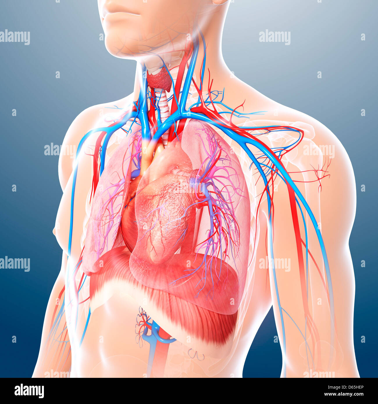
Chest Anatomy Artwork Stock Photo Alamy from c8.alamy.com The forehead is referred to as the frontal region. See chest anatomy stock video clips. Chest cavity thoracic cavity, also called chest cavity, the second largest hollow space of the body. Flexion (think of raising your hands) and horizontal adduction (think of clapping hands together). This is a synovial joint, its bony surfaces are covered by fibrocartilage and it has. The upper chest has two main functions: Your abdomen contains the digestive and urinary systems. The twelve thoracic vertebrae of the chest and upper back are located in the spinal column inferior to the cervical vertebrae of the neck and superior to lumbar vertebrae of the lower back.
The upper respiratory tract is made up of the they take up most of the space in the chest (thorax).
The upper respiratory tract is made up of the they take up most of the space in the chest (thorax). I lost my job and my medical coverage and have been taking prednisone, 15mg per day, as a stop gap. Related posts of anatomy of the chest area. Related posts of anatomy of the chest area. The axilla is the name given to an area that lies underneath the glenohumeral joint, at the junction of the upper limb and the thorax.it is a passageway by which neurovascular and muscular structures can enter and leave the upper limb. Upper can be felt in upper parts of chest, lower is in back. Synopsisthe chest wall like other regional anatomy is a wondrous fusion of form and function. The pectoral region is located on the anterior chest wall. Enlargement will result in bulging of the. Anatomy of the chest area. The chest anatomy includes the pectoralis major, pectoralis minor and the serratus anterior. Your abdomen contains the digestive and urinary systems. / upper back pain and chest pain can occur together.
The diaphragm forms the upper surface of the abdomen. Anatomy of the chest area. Related posts of anatomy of the chest area anatomy of shoulder. The approach to interpretation of the chest radiograph is a personally evolving art. Upper division of left superior lobar bronchus.
Figure Anatomy Of The Thymus Gland Pdq Cancer Information Summaries Ncbi Bookshelf from www.ncbi.nlm.nih.gov Anatomy of shoulder 12 photos of the anatomy of shoulder anatomy of nerves in shoulder, anatomy of posterior shoulder dislocation, anatomy of right shoulder, anatomy of shoulder labrum tear, anatomy of the shoulder games, human anatomy, anatomy of nerves in shoulder, anatomy of posterior shoulder dislocation, anatomy of right. Huge collection, amazing choice, 100+ million high quality, affordable rf and rm images. The upper chest has two main functions: Upper can be felt in upper parts of chest, lower is in back. Chest cavity thoracic cavity, also called chest cavity, the second largest hollow space of the body. This page provides an overview of the chest muscle group. The pec major) is the one that commands the most real estate. Thus, the right side of the image is the patient's left.
Dermatomes of the upper limbs are innervated by spinal nerves c5.
The epidermis is the outermost layer that provides a protective, waterproof seal over the body. Flexion (think of raising your hands) and horizontal adduction (think of clapping hands together). The circulatory system does most of. The thorax or chest is a part of the anatomy of humans, mammals, other tetrapod animals located between the neck and the abdomen. The forehead is referred to as the frontal region. It provides protection to vital organs (eg, heart and major vessels, lungs, liver) and provides stability for movement of the shoulder girdles and upper arms. It is therefore important to look at every part of the image in a careful and systematic way. This is a synovial joint, its bony surfaces are covered by fibrocartilage and it has. The spinal cord represents the cns in the thorax and serves as the vital link between the brain and the body. Related posts of anatomy of the chest area anatomy of shoulder. I lost my job and my medical coverage and have been taking prednisone, 15mg per day, as a stop gap. Of the two chest muscles, the pectoralis major (a.k.a. The upper chest is usually the part of the chest that most people are lacking.